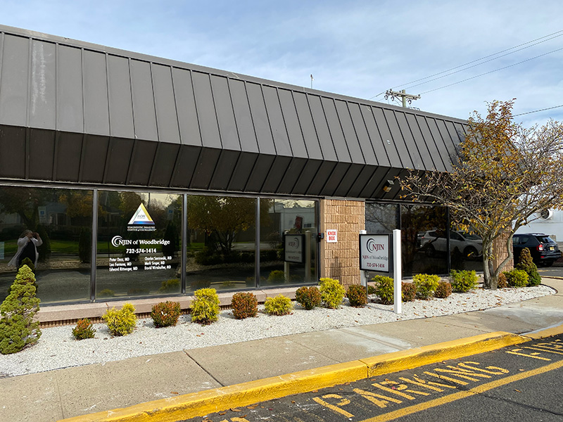
MRI, short for Magnetic Resonance Imaging, is a radiation-free scanning technique that uses radio waves and magnetic fields to produce detailed three-dimensional images of the body. MRI can be used to identify or precisely locate an injury or abnormality, to scan for developing problems or analyze damage from previous trauma, and to aid in the planning of surgery.
The MRI procedure lasts approximately 20-40 minutes, depending on the type of scan. The patient lies on a table that slides into the MRI unit. During the scan, the patient may listen to music or the radio via headphones that we will provide. Depending on the type of exam, intravenous contrast may be administered.
Patients who are particularly claustrophobic should discuss this with their referring physician and our office staff when scheduling their exam.
Click here for more information on MRI
Ultrasound or sonography produces images using high-frequency sound waves. It is especially useful for examining the breasts, bladder, thyroid, abdominal organs and male and female reproductive organs, and for obtaining images of fetuses in the womb. In addition, ultrasound is often used as a real-time guide during needle biopsies for the precise sampling of tissue.
During the procedure, the patient is positioned on an examination table and a warm, clear gel is applied to the area being examined. A transducer is then firmly pressed against the skin and swept back and forth to obtain the image.
DRCMRI was one of the first 1,000 vascular laboratories in the U.S., Canada and Puerto Rico to be granted accreditation by the Intersocietal Commission for the Accreditation for Vascular Labs.
Click here for more information on Ultrasound
X-ray imaging, also called radiography, is a fast and easy way to identify and diagnose bone injuries and disorders such as arthritis, cancer, pneumonia, fractures and infections. It can also be used to aid orthopedic surgeons in proper alignment of factures, and can aid in the detection and diagnosis of abnormalities in the chest, including the heart and lungs. X-rays may be followed up with MRI, PET, CT, or ultrasound imaging if further testing is needed.
Click here for more information on X-rays
back to top
Computed tomography (CT), or CAT scan, uses low doses of X-rays to obtain images of the body. CT scans are highly useful for examining injuries and abnormalities, guiding needle biopsies and aiding in surgical preparation.
The patient is positioned on a table on his or her back, side or stomach, and may be provided with pillows for comfortable support. The table moves very slowly through the doughnut-shaped CT scanner. The X-ray beam inside the CT unit spirals slowly around the patient on all sides, creating 360-degree images or "slices" of the area being examined. As the patient moves through the unit, many slices are captured. The images are then combined to produce a highly detailed, three-dimensional digital image.
Click here for more information on Computed Tomography, also known as CAT Scan
Mammography is an X-ray exam used to obtain images of the breast. It is a highly useful tool in the early detection of breast cancer because it may show an abnormality in breast tissue before the patient or doctor can feel it. Screening and diagnostic mammography can aid in the detection and diagnosis of breast diseases, lumps, cysts and benign and malignant tumors.
During the procedure, the breast is placed on a small platform and compressed with a paddle while it is exposed to a very low dose of radiation. Images are then displayed on a computer screen or printed on special film. Yearly mammograms are recommended for women over 40. Those who are at increased risk of developing breast cancer should consult with their physician about when to start having mammograms.
DRCMRI is ACR & FDA certified in mammography.
Mammographic studies performed at our Center are analyzed by a computed aided detection system to increase sensitivity in detecting cancers.
Click here for more information on Mammograms
back to top
All of our screening mammograms are read by our specially-trained radiologists using the iCAD Second Look system. The Second Look 300 is a sophisticated computer-aided detection system (CAD) that helps lower the number of overlooked breast tumors and cancers. After the radiologist evaluates a patient's mammogram results, the Second Look system scans the same images looking for suspicious masses, serving as an efficient "second pair of eyes."
This sophisticated computer-aided detection system aids in the review of the mammograms. The radiologist reads the mammogram in conjunction with this computer aided diagnostic tool. For more information, please visit iCAD’s website at www.icadmed.com.
DEXA (Dual-Energy X-ray Absorptiometry) is an enhanced X-ray image of the skeleton that provides the most accurate measurements of bone density available. Bone density tests are used to determine whether patients have osteoporosis, a condition in which bones are weakened and fracture easily.
During the procedure, a beam of low-dose X-rays is targeted at the area, usually the lower spine and hips. The energy is measured as it is absorbed by soft tissue and by bone, and computer software then processes the gathered information. Exams are painless and take 10-30 minutes.
Click here for more information on DEXA
|